
In the immediate future, healthcare is headed in the direction of early disease detection and precision diagnosis. As selected cancer types can now be accurately assessed using molecular imaging (PET/CT and SPECT/CT), early accurate staging and treatment can now be guided by using services such as those of Theranostics & Nuclear Medicine in Bangalore that offer specialized molecular imaging and targeted radionuclide therapies. In most cases, these cutting-edge technologies are best utilized in conjunction with an accurate early clinical diagnosis rather than as a replacement for it.
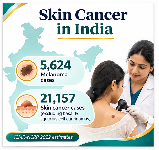
Number of new cancer cases in India, as estimated by the National Cancer Registry Programme of the Indian Council of Medical Research (ICMR-NCRP) for 2022 – 1.46M. Number of new cutaneous melanoma cases in India, as estimated by the National Cancer Registry Programme of the Indian Council of Medical Research (ICMR-NCRP) for 2022 – 5,624.
The numbers for melanoma, although small in terms of the overall cancer incidence in India, do warrant timely detection and accurate staging. However, it is to be noted that other skin cancers like basal cell carcinoma and cutaneous squamous cell carcinoma are reported differently by cancer registries.
There is a major difference between routine skin care and investigation for cancer. While the majority of skin lesions, such as rashes and pigmentation changes, are benign and don’t require advanced imaging, there are occasions when a suspicious lesion changes and requires evaluation by a specialist using dermoscopy and potentially a biopsy to establish a diagnosis and ensure appropriate treatment.
Referral pathways would thus need to be established to connect a dermatologist to a pathologist, an oncologist, and a nuclear medicine department. The dermatological consultation may form the first step in this process. A Dermatologist in Andheri West can be approached for a dermatological consultation.
1. India’s Skin Cancer Burden Requires Better Recognition
Skin cancer is an important but frequently misunderstood aspect of the overall cancer burden in India.
The estimated number of new cases of skin cancers not including basal and squamous cell carcinomas is 21,157 in the ICMR-NCRP reporting category for 2022. Of these cases, 5,624 were cutaneous melanomas.
The figures above do not account for basal and squamous cell carcinomas (as they were reported in a different category for skin cancer excluding these two types of skin cancers).
A report of cancer incidence from 43 Indian cancer registries across the country for the year 2025 also stressed the importance of consistent cancer classification and cancer surveillance through registries.
These statistics need to be interpreted carefully for estimating cancer prevalence in the population for planning cancer awareness programs, for teaching dermatological and surgical postgraduates and for making required health care resources available.
With accurate numbers of skin cancer in a population, awareness programs, more dermatological education for medical students, and health care resources can be appropriately allocated.
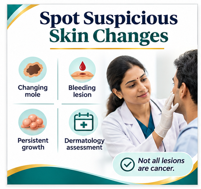
Persistent skin lesions, unexplained bleeding, changes to pre-existing moles or novel lesions that are suspected to be malignant need to be evaluated by a doctor.
2. Dermatology Is the First Step in Identifying Suspicious Lesions
Dermatology is the first step in identifying suspicious skin lesions. Initially, these need to be distinguished from common inflammatory, infectious, and pigmentary skin disorders.
An examination of a lesion for features such as size, color, border, and symmetry, and how it has changed over time.
Dermoscopy increases the depth of information collected from the skin and reveals structures below the surface of the skin.
However, dermoscopy does not replace histopathological examination when biopsy is clinically indicated.
When deciding on a suitable biopsy technique for suspected skin cancer, consideration must be given to the clinical diagnosis, location of the lesion, and relevant clinical guidelines.
Similarly, a clinically suspicious melanoma needs to be evaluated through an appropriate diagnostic pathway instead of considering it only as an unusual pigmentation lesion to be treated with a cream.
Clinically important lesions should be evaluated through an evidence-based diagnostic approach, not by considering them as cosmetic changes of normal skin.
3. Molecular Imaging Has a Defined Role in Cancer Staging
Nuclear medicine uses radiopharmaceuticals to examine specific biological processes.
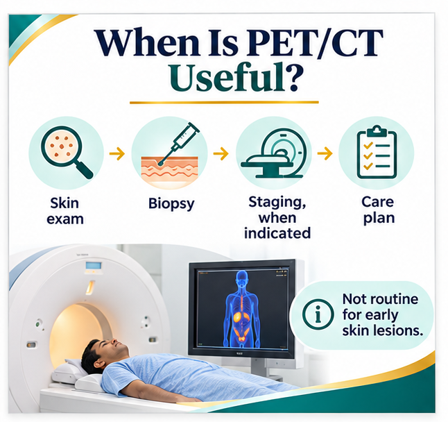
Nuclear medicine uses the appropriately selected radiopharmaceutical for the assessment of specific biological processes and therefore has a defined role in cancer staging using PET/CT for the assessment of cancers and their spread.
The demand of the different tumors, however, varies greatly.
The European Society for Medical Oncology (ESMO) guidelines on PET and CT for staging in cancer patients include a section on staging patients with higher risk or advanced melanoma and options for staging using other imaging modalities.
Routine PET/CT is not indicated for all patients with early-stage melanoma or a solitary indeterminate-looking incompletely biopsied mole.
Imaging can assess involvement of lymph nodes as well as of distant sites of metastases and also of treatment response in patients with advanced-stage melanoma and in specific situations in patients with higher-risk melanoma.
It is the whole multidisciplinary team’s decision after careful consideration of the individual’s circumstances.
Molecular imaging of tumors can help select the right investigation for the right patient and thus avoid increasing the number of scans performed.
4. Theranostics Represents a Broader Precision Oncology Opportunity
Theranostics combines molecular targeting with diagnostic imaging and, where appropriate, targeted radionuclide treatment.
In particular, the approach of theranostics is useful to identify biological targets and to check if a corresponding therapeutic radiopharmaceutical is available for the patient.
Among others, selected thyroid cancers, neuroendocrine tumors and prostate cancer are established applications of theranostics.
For example, radioactive iodine is already used to treat differentiated thyroid cancers, whereas the radiopharmaceutical lutetium-177 is applied in selected cases of prostate cancer and neuroendocrine tumors.
Note, though, that radioactive iodine is already a well-established treatment for selected cases of differentiated thyroid cancer and not a routine treatment for cutaneous melanoma or other dermatological tumors.
The interest of theranostics for us is to illustrate how selected molecular information can be used for individualized oncology.
One should also recall that India’s healthcare system can greatly benefit from incorporating advanced imaging and nuclear medicine in appropriate cancer care pathways.
5. Early Diagnosis and Advanced Technology Must Be Balanced
Early Diagnosis and Advanced Technology Must Be Balanced – An inclusive approach is required to improve cancer detection and treatment across India.
Advanced molecular imaging can be used to support complex treatment decisions. However, the majority of initial cancer patients will require simple evaluation by clinical examination, with the taking of a suitable biopsy for accurate and proper pathological evaluation.
Unnecessary investigations will increase the cost of cancer care, create undue anxiety, and result in avoidable harm.
On the other hand, referring a suspicious-looking lesion for investigations much later can make a complicated diagnosis and its treatment too.
The aim of healthcare therefore should be a structured pathway for primary assessment, dermatology, pathology and, in the end, cancer therapy.
For the right patient, Nuclear Medicine investigations can provide useful information after a diagnosis has been made.
These investigations have to be correlated with the individual tumor risk and have to be performed according to the current treatment guidelines.
6. India’s Next Priority Is Coordinated Cancer Diagnostics
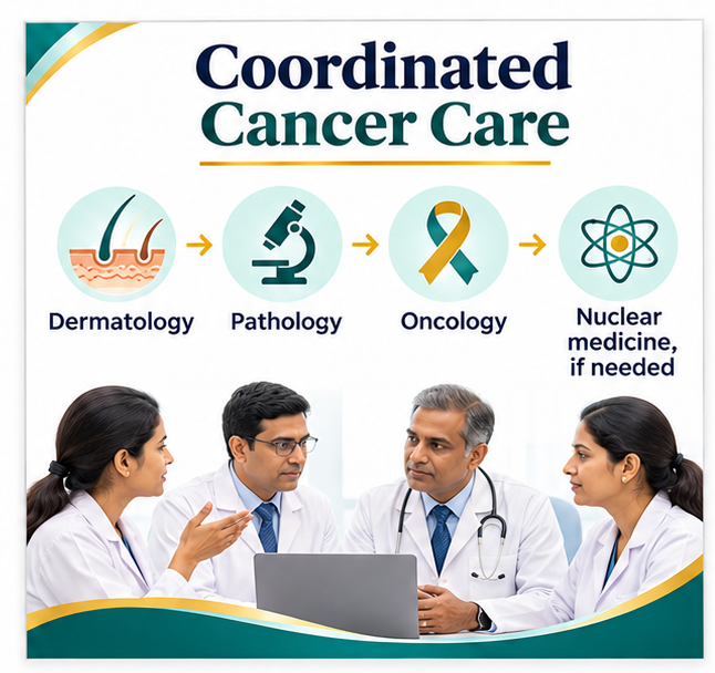
It is therefore very important for cancer development to focus on the integration of various specialties of cancer for the development of cancer care in India rather than focusing on individual cancer diagnostic techniques.
A dermatologist would identify a suspicious change in the skin and then do the initial investigations to confirm a diagnosis of cancer.
Pathologists determine tissue diagnosis and provide relevant clinicopathological information for treatment.
Oncology teams assess the stage of cancer and decide on the appropriate treatment.
Nuclear medicine specialists like to contribute by doing a molecular imaging study or even a targeted treatment when clinically indicated.
The organized model of healthcare will lead to more accurate diagnosis and less duplication of diagnostic investigations.
Ultimately, health care outcomes have to be assessed on the basis of the time taken to arrive at a correct diagnosis, choosing the right form of treatment, ensuring the patient’s safety, and following up on him or her for a long time to come.
Conclusion
Cancer is emerging as a growing health problem in India. Hence, there is a growing need to correlate a basic clinical evaluation with advanced diagnostic modalities for its proper management.
Dermatology and molecular imaging have different but complementary roles to play in certain cancer pathways.
While the majority of skin conditions can be evaluated by dermatology, there are some advanced cancers that could benefit from specialist nuclear medicine imaging and a multi-disciplinary approach.
A new milestone for cancer diagnosis in India is not in developing newer technologies for cancer diagnosis but in putting them to effective use for delivering accurate cancer diagnosis followed by proper investigations and the most effective treatment at the appropriate time in the cancer patient’s continuum of care.